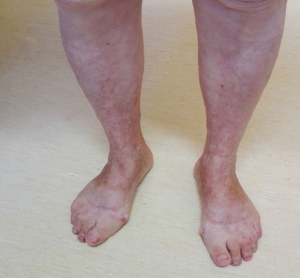
What should I know about PPD and its causes?
Pigmented purpuric dermatosis (PPD) is a rare benign and chronic disease caused by leaky small blood vessels called capillaries in the lower limbs. This causes extravasated red blood cells and hemosiderin to deposit in the tissues under the skin eventually leading to the skin red, purple or brown discoloration. Patients with pigmented purpuric dermatosis do not have any underlying varicose veins, however it is important that they are seen by vein specialist, as their skin changes should be differentiated from other similar vascular conditions such as venous eczema or hemosiderin skin staining. These cases may present as red or brown skin discoloration usually around the ankles, secondary to underlying varicose veins or hidden varicose veins. In these patients chronic inflammation causes skin damage and patchy stains as a result of blood break down and leaky little blood vessels. Exercises, capillary fragility, some medications as well as immunity factors can also play a role leading to red blood cells extravasation into the skin with characteristic purpuric color. Pigmented purpuric dermatosis is often asymptomatic but sometimes can be associated with itchines. Most patients present with skin changes on lower legs spontaneously without known cause. It is harmless skin condition not related to any major underlying disease or any clotting disorders but it can flare up and persist for years. It is important that patients presenting red, purple or brown skin stains see a venous specialist, even if varicose veins are not seen on your legs. Hidden varicose veins may cause inflammation of the skin and if left untreated, eventually lead to leg ulcer formation.
How can PPD be diagnosed ?
Pigmented purpuric dermatosis is usually diagnosed based on clinical presentation. However to rule out other possible causes, such as incompetent truncal veins or hidden varicose veins, comprehensive Duplex scan study with proper assessment of superficial and deep veins in leg is required for differential purpose. Careful assessment of perforators performed by specialist sonographer during the study is of most importance, as patient presenting with skin changes at the ankle often have incompetent perforating veins. Fluid collection with soft tissue swelling found during examination can be an additional important diagnostic factor showing the evidence of leaking veins with edema.
In patients with pigmented purpuric dermatosis Duplex scan will often appear normal, however if incompetent veins are found during scanning, they can be successfully treated. In case of normal ultrasound findings with no venous underlying disease, skin biopsy can be helpful. A very small sample of skin is removed using a punch biopsy and examined under microscope to confirm the final diagnosis. Blood tests are usually a useful tool to rule out blood clotting disorders or thrombocytopenia. If an allergic etiology is considered, patch testing can be also performed. Making a correct diagnosis is imperative to reassure patients and to avoid further costly referrals and unnecessary treatment.


